

Consultants’ Comments
- “I love being able to mark my own margins – I know right where they are.”
- “The Optispray (Sirona) is the easiest way to powder the tooth.”
- “I can’t tell that a restoration is there.”
- “Patients are impressed by the technology and enjoy having only a single visit.”
- “Take the time necessary to get good images, and the rest is easy.”
- “Lost some occlusal anatomy during adjustment.”
Description
CEREC 3 is a CAD/CAM system for in-office designing and milling of ceramic restorations. The complete system consists of an integrated computer/monitor/keyboard/camera, which is the hardware basis for the 3D software. The chairside imaging unit communicates through either a wired or a wireless connection to the milling unit that can be placed anywhere in the office. The camera utilizes red light to capture images of powdered tooth surfaces. The powder is mandatory for proper imaging. On-screen prompts guide the user through the various steps of margin marking, virtual die trimming and restoration design. Biogeneric (Sirona) software utilizes data from the patient’s tooth, images of a bite registration, and a library of tooth anatomy to create a digital proposal of the desired restoration. Design tools allow customization of the restoration, and a multi-color display shows strength of occlusal and interproximal contact information. The CEREC MC XL mill accepts ceramic or composite blocks of various sizes; the computer determines the minimum size compatible with each restoration. Milling is accomplished by two diamond burs under constant water spray. The milling chamber is illuminated for viewing, and the menu displays current status of the milling process.
Clinical Evaluation Protocol
- CEREC 3 was evaluated by Editors of THE DENTAL ADVISOR during a 12-month period.
- 161 restorations were designed and milled with the CEREC 3 system.
- Type of restoration, ceramic material and cement were noted.
- 28 restorations were available for evaluation of clinical performance up to 12 months after placement.
- Restorations were evaluated on a 1-5 rating scale: 1 = poor, 2 = fair, 3 = good, 4 = very good, 5 = excellent.
Placement
Obtaining good results starts with proper tooth preparation, meticulous attention to soft tissue retraction, uniform powdering, and accurate imaging. As users gain experience and familiarity with the system, the process can be carried out more smoothly and required less time. The majority of restorations fabricated were partial-coverage restorations, including inlays, onlays and ¾ crowns (Figure 1).
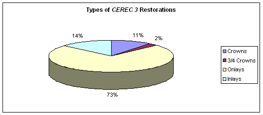 Figure 1.
Feldspathic ceramic (CEREC Blocks by VITA) was used for all of the early restorations. Later in the evaluation, IPS Empress CAD and IPS e.Max CAD (Ivoclar Vivadent) ceramics were used (Figure 2). The ceramics are available in high, medium and low translucency, as well as polychromatic blocks with a gradient of high to low translucency within the block. Generally, highly translucent blocks were selected for intracoronal restorations. The multi-colored blocks enhance the natural appearance of full and ¾ crowns.
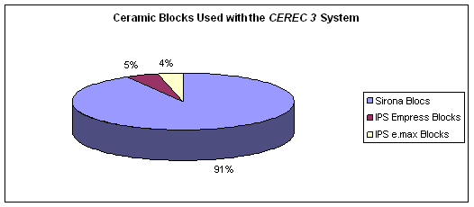 Figure 2.
The accuracy of the imaging was exhibited by the excellent fit of the milled restorations. Adjustment of proximal contacts or occlusion is sometimes necessary; some restorations seat with no adjustment. The variability in need for adjustment is a result of many factors including the level of experience of the dentist or assistant doing the design, tooth preparation, parameter settings, and sprue location. All restorations were clinically acceptable and were bonded with a variety of resin cements (Figure 3).
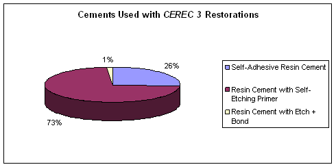 Figure 3.
Ceramic restorations milled with the CEREC MC XL were rated excellent for fit to the tooth (Figure 4). Proper tooth preparation, especially tapered walls and rounded corners, is the foundation for milled restorations that seat with no interference. Likewise, with good soft tissue retraction and accurate margin marking during the design phase, restoration margins in the mouth were observed to be excellent. The use of highly translucent ceramic and translucent cement enhances the esthetics of inlays and onlays, allowing visual blending of the restorative material with the tooth enamel. Anatomy is dependent upon the operator producing the design and is always limited by the milling burs, which do not have the ability to create deep occlusal grooves. After milling, the occlusal anatomy can be refined with a fine diamond bur in a high-speed handpiece and then polished.
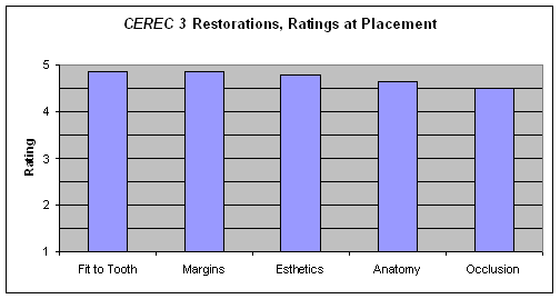 Figure 4.
Recall
Twenty-eight CEREC 3 restorations were evaluated at up to 12 months after placement. No restorations failed (defined as de-bonding or fractures that required replacement). Three of the restorations (10%) had minor chips on the occlusal surfaces; none of these required repair (Figure 5). Most of the intracoronal restorations were finished with ceramic polishers, while most of the crowns were glazed and fired. The glazed ceramic maintained its gloss, and the polished ceramic was sufficiently glossy in most cases.
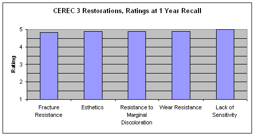 Figure 5.
Summary
CEREC 3 was used to fabricate 161 restorations, 28 of which were evaluated at 6-12 months after placement. The system is an effective and efficient means of delivering an indirect restoration in a single patient visit. At one-year recall, the majority of CEREC 3 restorations received excellent ratings. No visible signs of wear were noted and nearly all restorations were resistant to staining at the cavosurface margins. In most cases the ceramic exhibited excellent blending with the surrounding enamel. No post-operative sensitivity was reported.
Editors’ Note:
The CEREC 3 is no longer available for purchase. The MC XL is available.


