
Consultants’ Comments
- “Can enhance visualization of distinct lesions.”
- “Camera is easy to attach.”
- “Non-invasive for the patient.”
- “Enhances the clinical exam.”
- “Magnification through the lens would be an advantage.”
Description
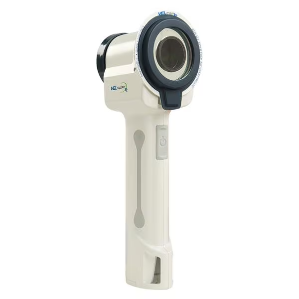
VELscope Vx is a hand-held device for use in conjunction with conventional intraoral and extraoral head and neck examinations. VELscope Vx provides clinicians with an adjunctive oral mucosal examination system that aids in the detection of a wide variety of oral conditions including cancerous and pre-cancerous lesions. The blue light emitted by VELscope Vx results in the fluorescence of oral tissues. The visualization of this fluorescence, including enhanced contrast between normal and abnormal tissue, is made possible through VELscope‘s proprietary filters. VELscope Vx may facilitate the discovery of oral mucosal abnormalities before they are visible under ordinary white light, which could lead to early diagnosis and treatment of oral disease. VELscope Vx may also be used by surgeons to help identify the appropriate margin for surgical excision. Photo-documentation can be accomplished by attaching a digital camera to VELscope Vx. The system includes the VELscope Vx handpiece with battery, charging cradle, power supply, power cord, 16 disposable VELcaps and a package of disposable plastic sheaths, patient safety glasses, 100 patient brochures, Quick Reference & Step by Step Examination Guides, and a training DVD. A Vx digital camera system (Canon) is separately available. VELscope Vx was used by 11 consultants in 200 uses. It received an 88% clinical rating.
Equipment Features
VELscope Vx has a simple design that is easy for dentists to incorporate into clinical use. The training DVD included with the system should be viewed by clinicians prior to use, as it does an effective job of reviewing instructions for use and interpretation of observations. With experience, consultants found that identifying abnormal areas of soft tissue became easier. Under VELscope Vx light, normal tissue fluoresces and appears green. Abnormal areas typically appear dark, and the user learns to discern characteristics that separate highly suspicious lesions from anatomical structures and vascular lesions. The VELscope Vx handpiece is cordless with a single button for operating the light. When viewing the mouth through VELscope Vx, objects may appear smaller (although, in fact, there is no demagnification); for dentists who are used to working with loupes, magnification through the filters would be helpful. When not in use, the light is stored on a cradle. The optional Vx digital camera system captures images, which can be printed for the patient’s record or as a tool for surgeons. Most consultants who evaluated VELscope Vx had been using manual and unaided visual exam techniques. Sixty-four percent found VELscope Vx to be better than unaided exam techniques for evaluating intraoral lesions and 27% found it to be equivalent. Sixty-four percent of consultants would switch to VELscope Vx,and 73% would recommend it to a colleague.
Clinical Tips
Dim the room lighting.